

Your dental X-ray, explained by a team of dentists.


Upload a panoramic, periapical or 3D scan — it goes to the specialist it concerns, and you get a description in plain language. Independent of your clinic, and without a diagnosis.
Real dentists describe your scan — not artificial intelligence.
Your scan is read by a practising dentist with decades of experience — the specialist whose field it actually concerns: surgery, endodontics, orthodontics, prosthetics or paediatric dentistry.
A scan isn't a picture. It's a story you have to know how to read.
"AI analysis" services find spots and draw boxes. A doctor sees the context: your age, old fillings, how the roots sit, what was done before — and knows what you should ask your dentist.
What an algorithm gives you
- Flags suspicious areas without explaining why they matter to you
- Doesn't answer the question you actually asked
- Can't tell an old filling from new decay without context
- A report of numbers and terms you still have to show someone
What a doctor gives you
- Reads the whole scan, with your age and complaint in mind
- Answers your question: "does it need to come out", "what is that shadow", "can it wait"
- Writes so you understand it the first time — no Latin, no abbreviations
- Independent of any clinic: has nothing to sell you
Three steps. Nothing extra.
Upload the scan
A file from the clinic, a scanned film or a photo of the screen — anything that can be examined. 3D scans go in as a ZIP archive.
≈ 1 minuteTell us about yourself
E-mail, age, sex and — if you like — what worries you. Your name is optional: the doctor needs the scan, not your passport.
≈ 1 minuteGet the report
The scan goes to a dentist of the right specialty. The plain-language report arrives by e-mail.
48 hSee how a scan is read
A description of your dental X-ray by an independent expert dentist.
The video loads from YouTube only after you press play.
This is what you receive
A real report on a real scan, written by a dentist. One A4 page, as a PDF, by e-mail.
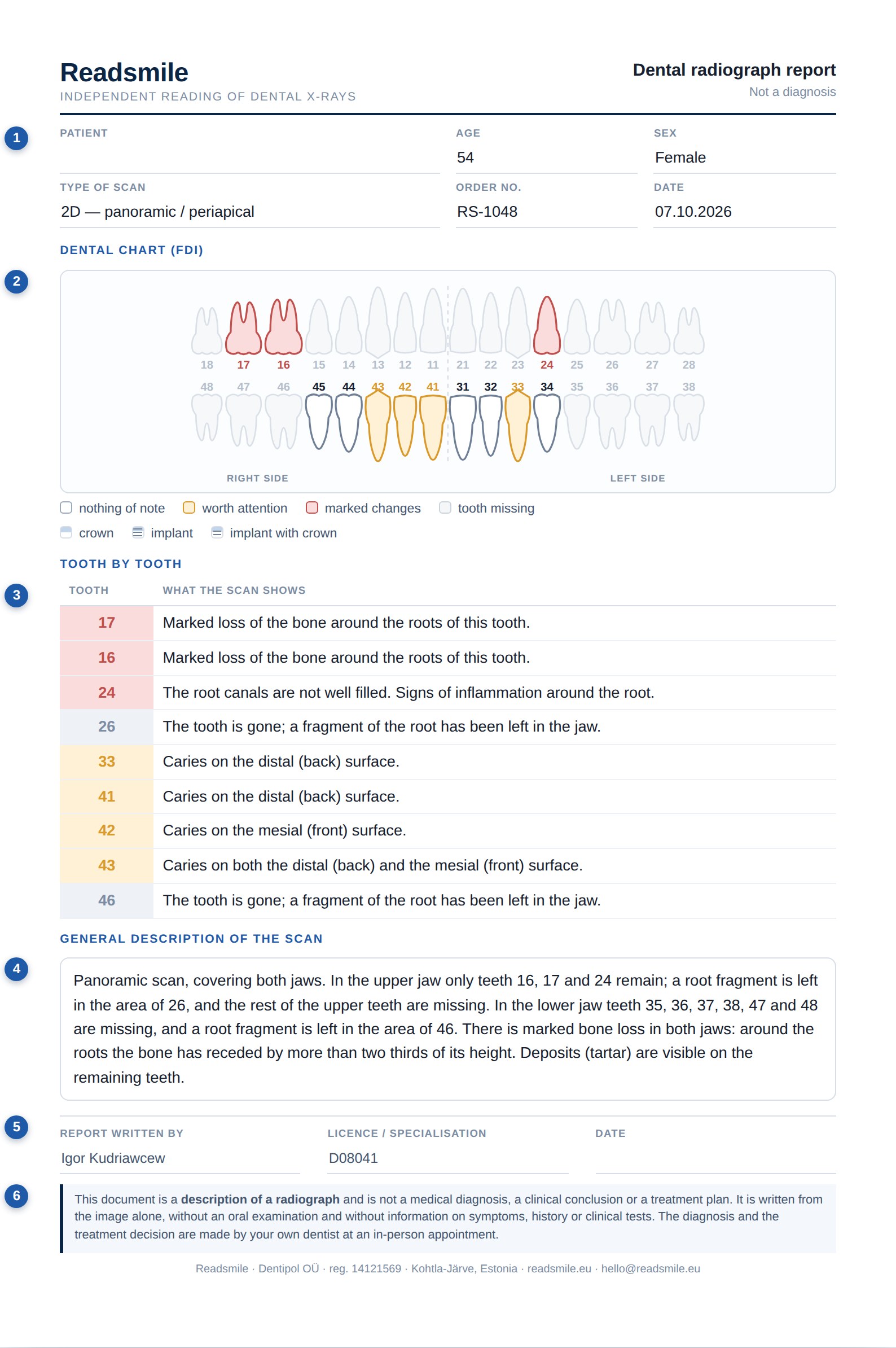
- 1Who and what was scanned
Age, sex, type of scan, order number and date. Your name is optional: the dentist needs the image, not your passport.
- 2The dental chart
Every tooth with its FDI number. Yellow is worth attention, red is marked changes, pale grey means the tooth is missing. Crowns and implants have their own symbols.
- 3Tooth by tooth
Each marked tooth gets its own line saying what the scan actually shows there, in plain language, with no Latin.
- 4The scan as a whole
Bone levels, sinuses, joints, previous treatment, anything that concerns the image in general rather than one tooth.
- 5Signed by the dentist
The name and the licence number of the doctor who read your scan. You always know who answered you.
- 6What this is not
A description, not a diagnosis. It does not replace an examination, and it says so on the page itself.
A genuine report on a real radiograph. The patient's name and all identifying details have been removed.
The doctors who read your scan
Every report is read and signed by a named dentist. We are not tied to any clinic and never refer you to “our own”.

Maxillofacial surgeon and prosthodontist. Reads panoramic and 3D scans and explains what they show in plain language.
- 1992–1997Department of maxillofacial and plastic surgery, Pavlov First St. Petersburg State Medical University
- 1997–1999Dr. Alperini clinic, Buenos Aires, Argentina
- 1999–2009Head of prosthetic dentistry, Polyclinic 24, St. Petersburg
- 2010–2014Department of implantology and prosthetic dentistry, Medical University of Gdańsk
- 2014–2017Ida-Viru Central Hospital, Estonia
- 2017–2020Hymylääkärit clinic, Helsinki
- 2020–nowPractice in Tallinn · Readsmile

General dentist, with further specialisations in endodontics (root canal treatment) and periodontology (gums).
- 2004–2020Chief dentist of the “Dobry Stomatolog” clinic network
- 2020–nowStomatoloogia Pluss dental clinic, Tallinn
- 2020–nowLead teacher, SP Training Center — dental assistant programme
The rest of the team stays unnamed — on purpose
Readsmile works with practising specialists in surgery, endodontics, orthodontics, prosthetics and paediatric dentistry. They read scans as independent experts and prefer not to be listed publicly: their opinion belongs to you, not to a clinic's marketing. Every report is signed by the doctor who wrote it, so you always know who answered you.
One scan. One dentist who read it. One clear answer.
- A plain-language report on any scan
- An answer to your question, if you asked one
- One follow-up question to the doctor — free
- Full refund if the scan cannot be read because of its quality

What people usually ask
Is this a diagnosis?
No. The doctor describes what is visible on the scan and explains what it may mean. The diagnosis and treatment plan are made by your dentist at an appointment — you arrive already knowing what to ask.
Where do I get my scan as a file?
Ask the clinic where it was taken — you are entitled to a copy. Usually it's a DICOM, JPG or PDF file sent by e-mail or on a USB stick. If you only have film, photograph it against a window.
Definitely not AI?
Definitely. A dentist opens the scan, reads it, writes the report in their own words and signs it. The report carries the name and specialty of the doctor who answered you.
What about 3D scans (CT, CBCT)?
Put the folder with the study into a ZIP archive and upload it as one file. If the clinic gave you a viewing link, paste it into the "What worries you?" field.
Who will see my scan?
Only the doctor who writes the report. Files are stored for 30 days and then deleted. Your name is optional.